
Level 8 Skelett der oberen Extremität I Propädeutik Makroanatomie… Memrise
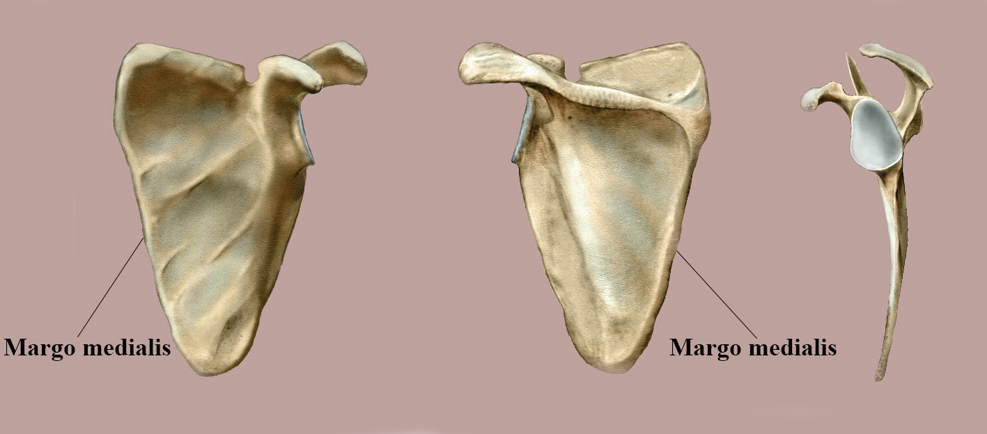
from the margo medialis of the scapula. We wish to communicate our tech-nique of a longitudinal osteotomy of the margo medialis for improved refixa-tion of the muscles. Patients and Methods: 5 patients with subscapular and one patient with a subrhomboid benign tumor were operated using this on technique.

Anatomy Standard Drawing Scapula posterior surface Latin labels AnatomyTOOL
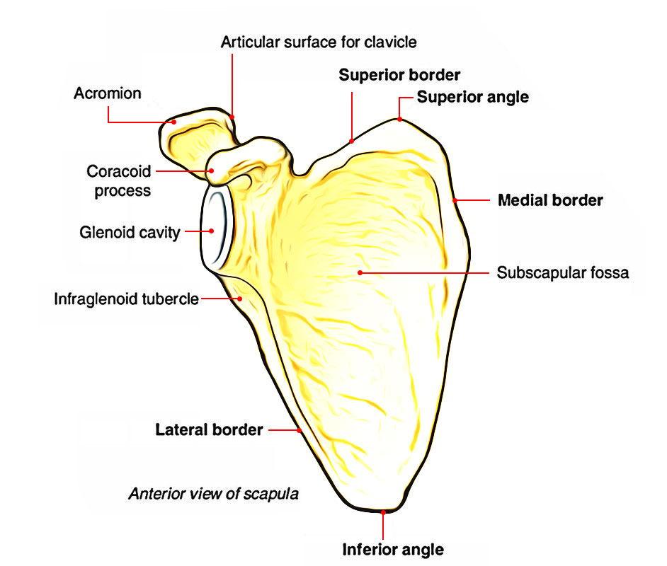
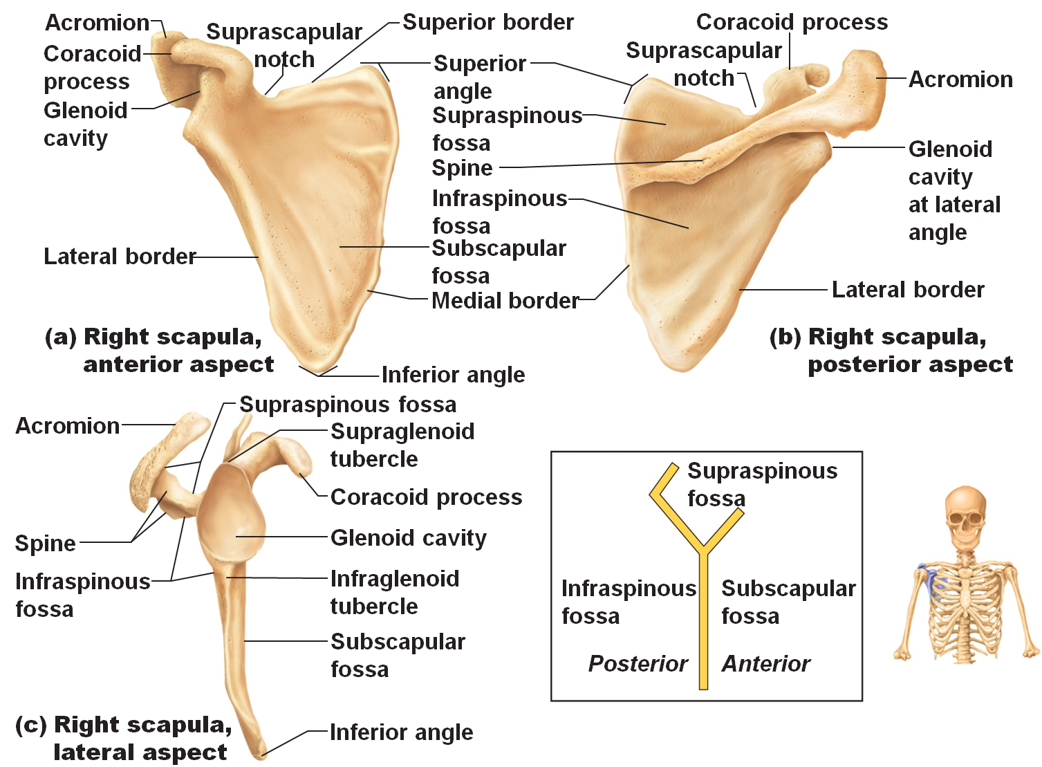
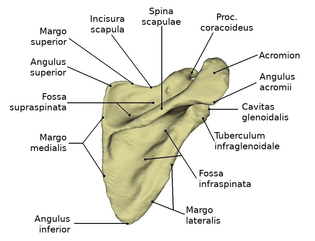
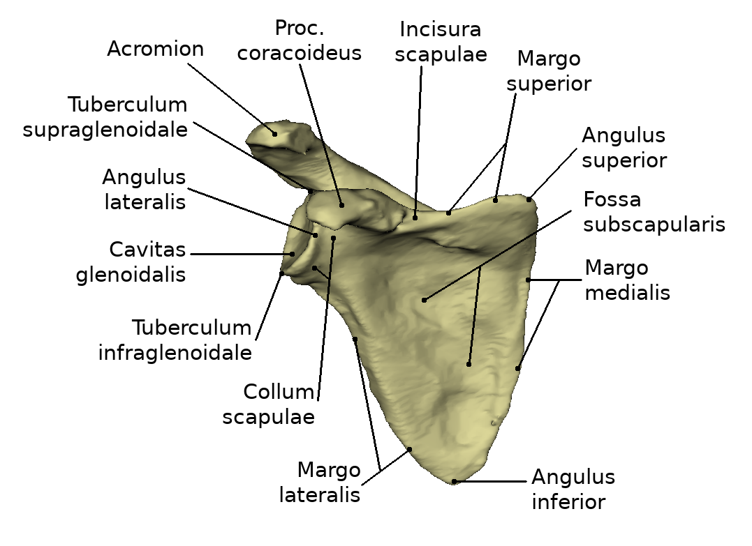
The scapula. Incisura scapulae Incisura scapulae Tuberculum infraglenoidale Tuberculum infraglenoidale Tuberculum supraglenoidale Tuberculum supraglenoidale Acromion Acromion Fossa subscapularis Fossa subscapularis Facies costalis; facies anterior Facies costalis; facies anterior Collum scapulae Collum scapulae Tuberculum infraglenoidale.

Shoulder blade Dornheim Anatomy
Bone Scapula Acromion Processus coracoideus Cavitas glenoidalis scapulae Margo lateralis scapulae Margo medialis scapulae Angulus inferior scapulae Fossa subscapularis Fossa infraspinata Spina scapulae Fossa supraspinata Incisura scapulae Margo superior scapulae

Scapulalopatica
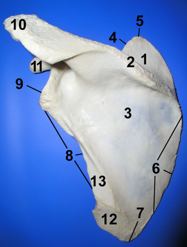
Flächen Linkes Schulterblatt, Facies dorsalis 1 Fossa supraspinata, 2 Spina scapulae, 3 Fossa infraspinata, 4 Margo superior, 5 Angulus superior, 6 Margo medialis, 7 Angulus inferior, 8 Margo lateralis, 9 Angulus lateralis, 10 Acromion, 11 Processus coracoideus, 12 Ursprungsfläche des Musculus teres major, 13 Ursprungsfläche des Musculus teres minor Linkes Schulterblatt, Facies ventralis

Two views of the Scapula Human anatomy and physiology, Medical knowledge, Yoga anatomy
3.3 Margo medialis (vertebralis) 4 Winkel 4.1 Angulus superior 4.2 Angulus inferior 4.3 Angulus lateralis 5 Prominente Strukturen 5.1 Spina scapulae 5.2 Processus coracoideus 5.3 Acromion 5.4 Cavitas glenoidalis 6 Entwicklung 7 Funktion 8 Klinik 9 Podcast 10 Bildquelle Definition Die Scapula bildet den hinteren Teil des knöchernen Schultergürtels.
Scapula (Shoulder Blade) Anatomy Earth's Lab
A second window on the medial border of the scapula can be made to aid reduction and/or to augment stability. Small (2.0-2.7 mm) plates in a 90° configuration on the lateral border and, if required, on the medial border are used.. The medial incision and precedent reduction of the the margo medialis is made neutralizing the medial.
medical freak THE SKELETON OF THE UPPER LIMB
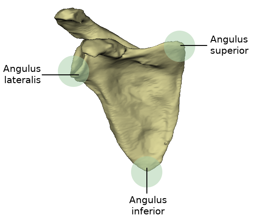
Medial border (margo medialis) Lateral border (margo lateralis) Superior border (margo superior) Suprascapular notch (incisura scapulae) The scapula has three angles: The superior angle (angulus superior) Superior angle (angulus superior) The inferior angle (angulus inferior) Inferior angle (angulus inferior)
Shoulder blade Dornheim Anatomy
Margo medialis scapulae är det latinska namnet på skulderbladets mediala kant. Synonymer margo vertebralis scapulae, "skulderbladets kant som vetter mot ryggraden. Margo medialis är skulderbladets längsta kant. Den sträcker sig från benets övre vinkel ( angulus superior) till dess nedre ( angulus inferior ).
A csontvázrendszer
See: medial border of forearm, medial border of foot, medial border of humerus, medial border of kidney, medial border of scapula, medial border of suprarenal gland, medial border of tibia. Synonym (s): margo medialis [TA], medial margin Farlex Partner Medical Dictionary © Farlex 2012 Want to thank TFD for its existence?
Schulterblatt
Like any triangle, the scapula consists of three borders: superior, lateral and medial. The superior border is the shortest and thinnest border of the three. The medial border is a thin border and runs parallel to the vertebral column and is therefore often called the vertebral border. The lateral border is often called the axillary border as it runs superolaterally towards the apex of the axilla.
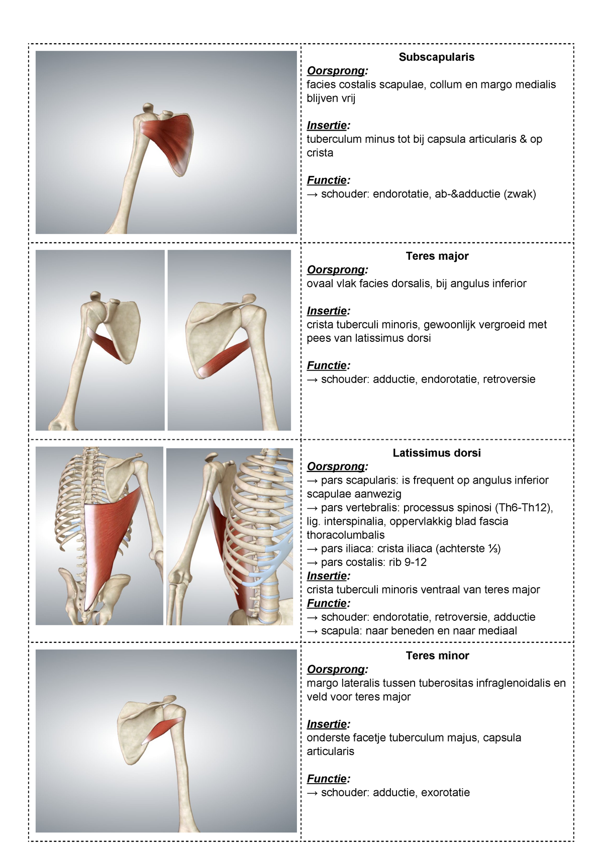
Spieren Subscapularis Oorsprong facies costalis scapulae, collum en margo medialis blijven
Margo medialis scapulae Description The medial border (vertebral border or vertebral margin) of scapula is the thin, medially located edge of the bone. It extends from the superior angle of the scapula to its inferior angle and is longest of three borders of the scapula, the other two being the lateral and superior borders.
Schulterblatt Dornheim Anatomy
Scientific Basis Occlusion Occipital Bone Parietal Bones Scapula Scapula, or shoulder blade is fixated to the axial skeleton solely via clavicle. Motions of the shoulder blade, to a great extent, facilitate the movements of the upper arm. Scapula in situ. Posterior oblique view.
Schulterblatt Dornheim Anatomy
Definition The medial border (vertebral border) is the longest of the three, and extends from the medial to the inferior angle. It is arched, intermediate in thickness between the superior and the axillary borders, and the portion of it above the spine forms an obtuse angle with the part below.

Question Wat is de scapula? Memory
Margo medialis scapulae. Margo lateralis scapulae. Angulus superior scapulae. Angulus inferior scapulae. Fossa infraspinata. Fossa supraspinata. Processus coracoideus. Uploaded by: rva Netherlands, Leiden - Leiden University Medical Center, Leiden University. Creator(s)/credit: Dr Eric Bauer, Biology professor.

Musculus serratus anterior Muscle anatomy, Body anatomy, Human muscle anatomy
The scapula is only connected to other bones via the clavicle. Latin labels. Image retrieved from Anatomy Standard, page Scapula. Anatomical structures in item: Scapula. Facies posterior scapulae. Angulus superior scapulae. Margo superior scapulae. Fossa supraspinata.

Scapula dan Clavicula Medical anatomy, Anatomy and physiology, Skeletal system anatomy
The scapula is a thick, flat bone lying on the thoracic wall that provides an attachment for three groups of muscles: intrinsic, extrinsic, and stabilizing and rotating muscles. The intrinsic muscles of the scapula include the muscles of the rotator cuff —the subscapularis, teres minor, supraspinatus, and infraspinatus. [3]