
Long Bone Diagram Labled Anatomy Test 3 at Westbrook High School
Clinical significance There are two congenital disorders of the long bones. In a disorder known as rachitis fetalis anularis the ends of the long bones (epiphyses) are enlarged. [2] Another disorder, rachitis fetalis micromelica, is a deficiency in the growth (as a shortness) of the bones. [2]
Long Bone Labeling Diagram / Long Bone Labeled Quizlet Microscopic
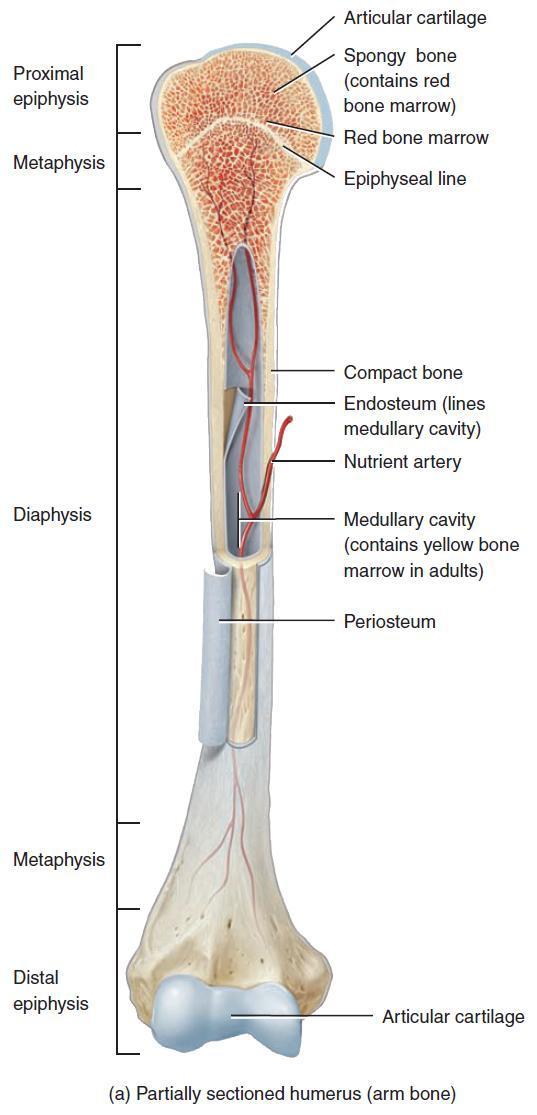
Description: Anatomy of long bones. A long bone (here: the femur) consists of epiphyses, metaphyses and a diaphysis (shaft). The epiphysial plate has been closed in this bone and has become the epiphyseal line after puberty. English labels. Case courtesy of Dr Matt Skalski, Radiopaedia.org. From the case rID: 29729 Anatomical structures in item:

Long Bone Labeled Epiphysis / Label A Long Bone
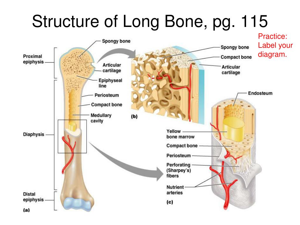
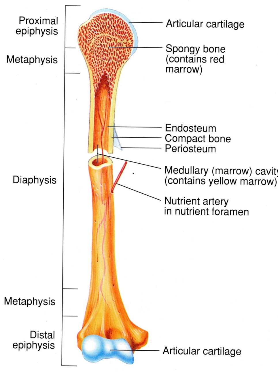
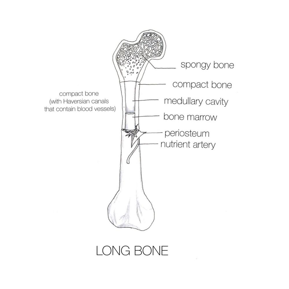
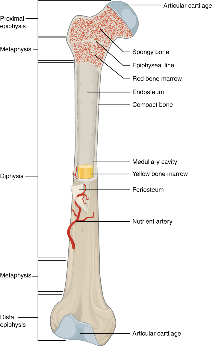
Anatomy of a Long Bone. A long bone has two parts: the diaphysis and the epiphysis. The diaphysis is the tubular shaft that runs between the proximal and distal ends of the bone. The hollow region in the diaphysis is called the medullary cavity, which is filled with yellow marrow. The walls of the diaphysis are composed of dense and hard.
Flashcards Diagrams StudyBlue
A long bone has two parts: the diaphysis and the epiphysis. The diaphysis is the tubular shaft that runs between the proximal and distal ends of the bone. The hollow region in the diaphysis is called the medullary cavity, which is filled with yellow marrow. The walls of the diaphysis are composed of dense and hard compact bone.

Long Bone Labeled Epiphyseal Plate Bone Growth And Development
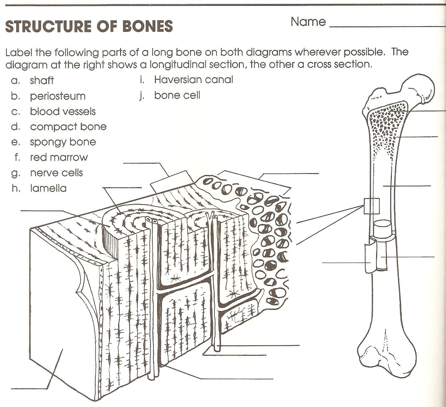
Label the Long Bone Structures 1. _____ 2.. bone marrow compact bone periosteum Sharpey's fibers arteries feeding the bone articular cartilage of the joint compact bone spongy bone ©Sheri Amsel www.exploringnature.org osteon or Haversian system (units of compact bone) epiphyseal line trabeculae spicules

Long Bone Labeling Worksheet Appendicular Skeleton Anatomy
L o n g B o n e L a b e l e d D i a g r a m 1. Diaphysis The central tubular shaft connects the two ends of the bone. Its walls are composed of dense and hard compact bone, forming an internal hollow region called the medullary cavity (as shown in the cross-section image above).

Anatomy Labeling and defining the long bone. Diagram Quizlet
Label a Long Bone Quiz Science » Image Quiz Label a Long Bone by mpurzycki +1 82,865 plays 13 questions ~30 sec English 13p 51 3.83 (you: not rated) Tries 13 [?] Last Played October 18, 2023 - 03:14 am There is a printable worksheet available for download here so you can take the quiz with pen and paper. Remaining 0 Correct 0 Wrong 0 Press play! 0%
Long Bone Labeling Worksheet Skeletal System Diagram Worksheet
The structure of a typical long bone - drawn, defined and discussed!The Human Body is a complex, amazing biological machine. 'Human Biology Explained' is a Y.
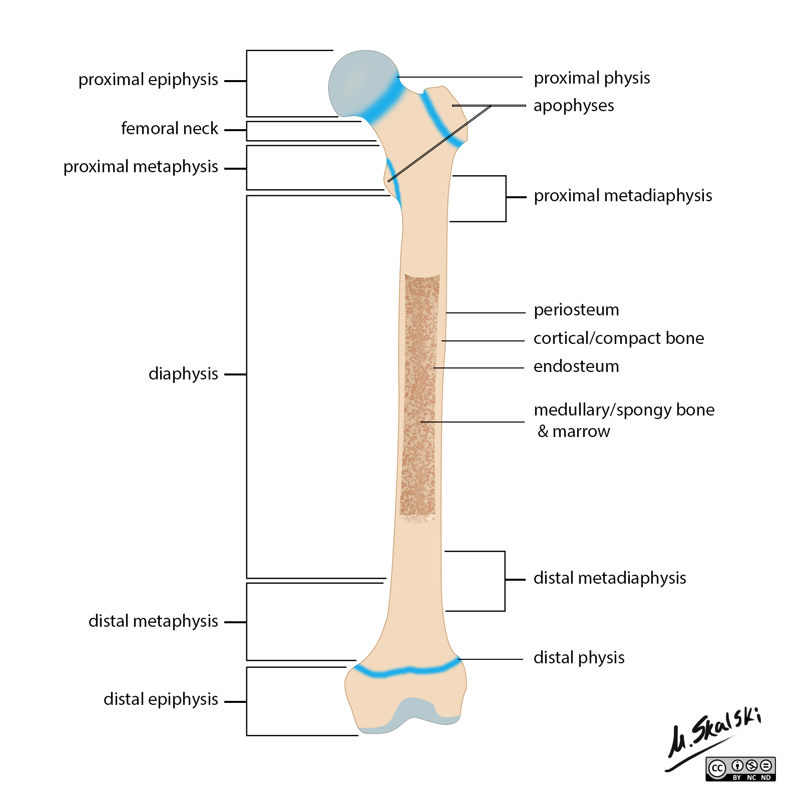
Radiopaedia Drawing Anatomy of long bones (femur) English labels
This article will provide detailed information on a long bone's gross and microscopic features with a labeled diagram. Again, I will provide the examples and functions of the long bones from different animals like dogs, cats, and ruminants. You will come to know the modified long bones and miniature long bones in different animals.
long bone structure diagram Quotes
High Quality, Long-lasting Labels for All Your Lab Labeling Needs. Order Now! Available in a Variety of Sizes and Materials. Perfect for Any Application. Shop Now!

Long Bone Labeled / Structure Of A Long Bone Human Anatomy And
Sesamoid bones vary in number and placement from person to person but are typically found in tendons associated with the feet, hands, and knees. The patellae (singular = patella) are the only sesamoid bones found in common with every person. Table 6.1 reviews bone classifications with their associated features, functions, and examples.

Long Bone Labeling / Long Bone Labeled Quizlet / A P I Compact Bone
This section will examine the gross anatomy of bone first and then move on to its histology. Gross Anatomy of Bones A long bone has two main regions: the diaphysis and the epiphysis ( Figure 6.3.1). The diaphysis is the hollow, tubular shaft that runs between the proximal and distal ends of the bone.
Long Bone Diagram Blank / Chapter 7 Skeletal System Ppt Download
Bone markings are invaluable to the identification of individual bones and bony pieces and aid in the understanding of functional and evolutionary anatomy. They are used by clinicians and surgeons, especially orthopedists, radiologists, forensic scientists, detectives, osteologists, and anatomists.
️Long Bone Diagram Worksheet Free Download Gmbar.co
In this video we discuss the parts of a long bone and some of the functions of each of those bone parts. We cover the diaphysis, the epiphysis, spongy and c.
Long bone Wikipedia
The skeletal system includes all of the bones and joints in the body. Each bone is a complex living organ that is made up of many cells, protein fibers, and minerals. The skeleton acts as a scaffold by providing support and protection for the soft tissues that make up the rest of the body. The skeletal system also provides attachment points for.

Long Bone Diagram 5 Best Images of Upper Limb Labeling Worksheet
The Structure of a Long Bone: Labelled Image Long bones are the most common bones found in the human body. They are composed mostly of compact bone, and are roughly cylindrical in shape with enlarged ends filled with spongy bone.